Femur Fracture Physical Therapy Protocol
This protocol details rehabilitation post-femur fracture, integrating surgical intervention, range of motion exercises, and weight-bearing progression for optimal recovery.
Femur fracture rehabilitation is a complex process demanding a multidisciplinary team – surgeons, physiotherapists, and specialists – to optimize patient outcomes. Early mobilization and tailored exercise regimens are crucial, addressing deficits stemming from the fracture and potential implant presence.
Post-operative care, particularly following intramedullary nailing, focuses on controlled weight-bearing and regaining functional mobility. This protocol aims to guide clinicians through phases of recovery, from initial pain management and edema control to advanced strengthening and return to activity, ensuring a safe and effective rehabilitation journey.
Understanding Femur Fracture Types
Femur fractures vary significantly, influencing rehabilitation approaches. Proximal femur fractures, often hip fractures, require careful consideration due to potential impact on mobility. Femoral shaft fractures, commonly treated with intramedullary nailing, necessitate weight-bearing protocols. Distal femur fractures, potentially comminuted, demand focused range of motion exercises. Understanding the fracture’s location, stability, and treatment method (surgical vs. non-operative) is paramount. Rehabilitation must be tailored to the specific fracture pattern, considering factors like patient age, health, and functional goals for optimal recovery.

Proximal Femur Fractures
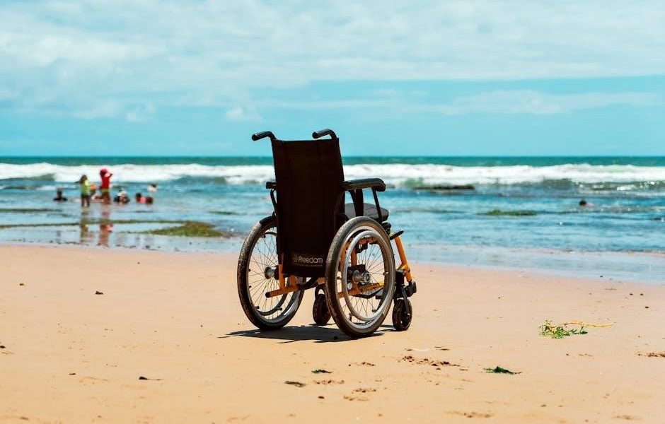
Proximal femur fractures, frequently occurring as hip fractures, often require early surgical intervention to facilitate recovery and pain management. Rehabilitation focuses on restoring hip mobility, strengthening surrounding musculature (glutes, quads, hamstrings), and regaining functional independence. Weight-bearing progression is carefully monitored post-intramedullary nailing. Proprioceptive training is crucial to improve balance and prevent falls. Physical therapy addresses gait abnormalities and prepares patients for activities like standing and walking, aiming to maximize functional capacity and quality of life post-fracture.
Femoral Shaft Fractures
Femoral shaft fractures commonly necessitate intramedullary nailing for stabilization, influencing rehabilitation protocols. Early mobilization, guided by weight-bearing restrictions determined post-surgery, is key. Physical therapy concentrates on restoring knee and hip range of motion, alongside strengthening exercises targeting quadriceps, hamstrings, and gluteal muscles. Addressing potential complications like delayed union requires tailored interventions. Proprioceptive exercises enhance stability, while gait training focuses on normalizing walking patterns. Careful monitoring and a multidisciplinary approach are vital for optimal functional outcomes.

Distal Femur Fractures
Distal femur fractures often require surgical intervention, potentially involving plates or intramedullary nails, dictating rehabilitation phases. Early physical therapy focuses on pain and edema management, alongside gentle range of motion exercises. Weight-bearing progression is carefully controlled, guided by fracture healing and implant stability. Strengthening exercises target quadriceps, hamstrings, and calf muscles, crucial for knee extension and stability. Proprioceptive training and functional exercises, like stair climbing, are incorporated as healing advances, aiming for full restoration of lower limb function.
Initial Post-Operative Phase (0-2 Weeks)
The initial phase (0-2 weeks) prioritizes pain management and edema control utilizing modalities like ice and elevation. Early range of motion exercises, focusing on ankle pumps, quad sets, and gentle hip/knee bends, prevent stiffness. Weight-bearing restrictions are strictly adhered to, guided by the surgeon’s protocol – often non-weight bearing or toe-touch weight bearing. Emphasis is placed on maintaining pulmonary toilet and promoting early, safe mobility within limitations. The goal is to minimize complications and prepare for progressive rehabilitation.
Pain Management and Edema Control
Effective pain management is crucial in the initial phase, often achieved through prescribed analgesics. Edema control utilizes elevation of the affected limb above the heart, frequent icing applications (20 minutes on, 20 minutes off), and gentle compression bandaging. Monitoring for signs of compartment syndrome is vital. Patient education regarding pain scales and reporting any increasing pain or swelling is paramount. These interventions optimize patient comfort and facilitate early mobilization, contributing to improved recovery outcomes.
Early Range of Motion Exercises
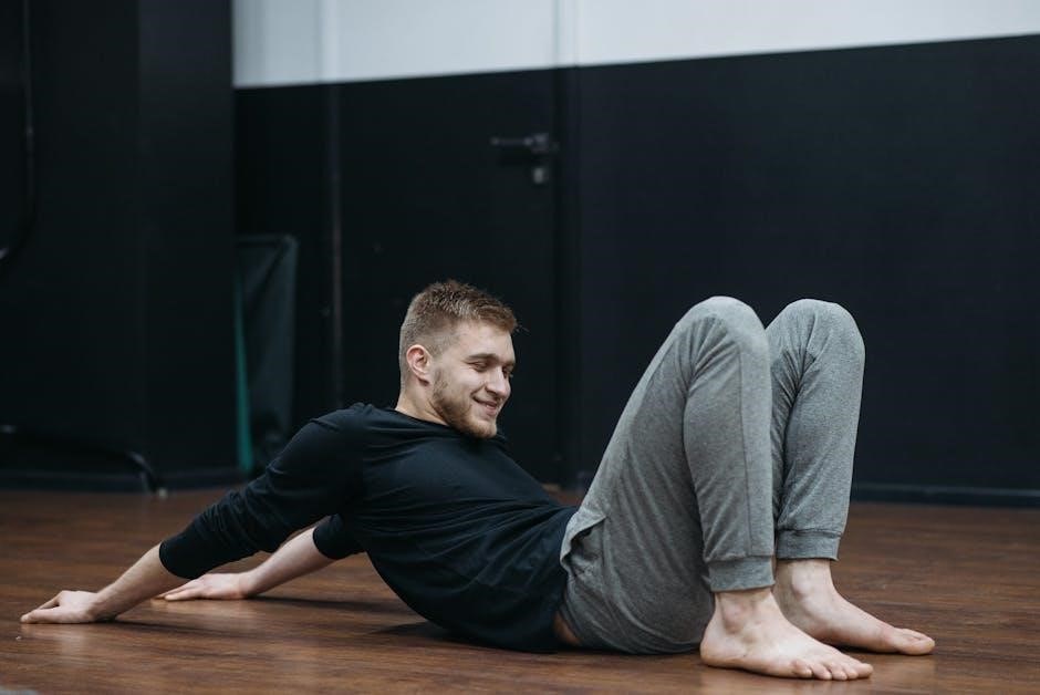
Gentle range of motion (ROM) exercises begin immediately post-operatively, focusing on unaffected joints to maintain function. Ankle pumps and circles are initiated to prevent deep vein thrombosis. Assisted knee flexion and extension, within pain-free limits, are introduced cautiously. Hip abduction and adduction exercises are also started gently. The goal is to prevent stiffness and maintain some degree of joint mobility without compromising fracture stability; These exercises are performed frequently throughout the day, guided by the physiotherapist.
Intermediate Rehabilitation Phase (2-6 Weeks)
This phase focuses on restoring functional movement and building strength. Weight-bearing progression, guided by the orthopedic surgeon, is a key component, potentially advancing from toe-touch to partial weight-bearing. Strengthening exercises target the quadriceps, hamstrings, and gluteal muscles, crucial for gait and stability. Proprioceptive training is initiated to improve balance and coordination. Early gait training with assistive devices, like a walker or crutches, begins, emphasizing proper mechanics. Pain management continues, and edema control remains a priority.
Weight-Bearing Progression
Weight-bearing advancement is carefully dictated by fracture stability and surgical fixation type, particularly with intramedullary nailing. Initially, toe-touch weight-bearing is permitted, progressing to partial weight-bearing (PWB) as tolerated, often around 2-4 weeks. Full weight-bearing (FWB) is typically achieved by 6-8 weeks, contingent on radiographic evidence of healing. Protocols vary; the surgeon’s guidance is paramount. Monitoring for pain and swelling is crucial throughout progression, adjusting the load as needed to prevent complications and promote bone healing.
Strengthening Exercises – Quadriceps, Hamstrings, Glutes
Progressive strengthening is vital for restoring lower extremity function. Early exercises focus on quadriceps sets, hamstring curls, and gluteal squeezes, performed isometrically. As strength improves, progress to short-arc quads, straight leg raises, and bridging exercises. Resistance bands and light weights are introduced gradually. Hamstring strengthening includes prone hip extensions and curls. Glute exercises progress to side-lying hip abduction and single-leg stance. Pain monitoring guides intensity; avoid overexertion to protect the healing fracture.
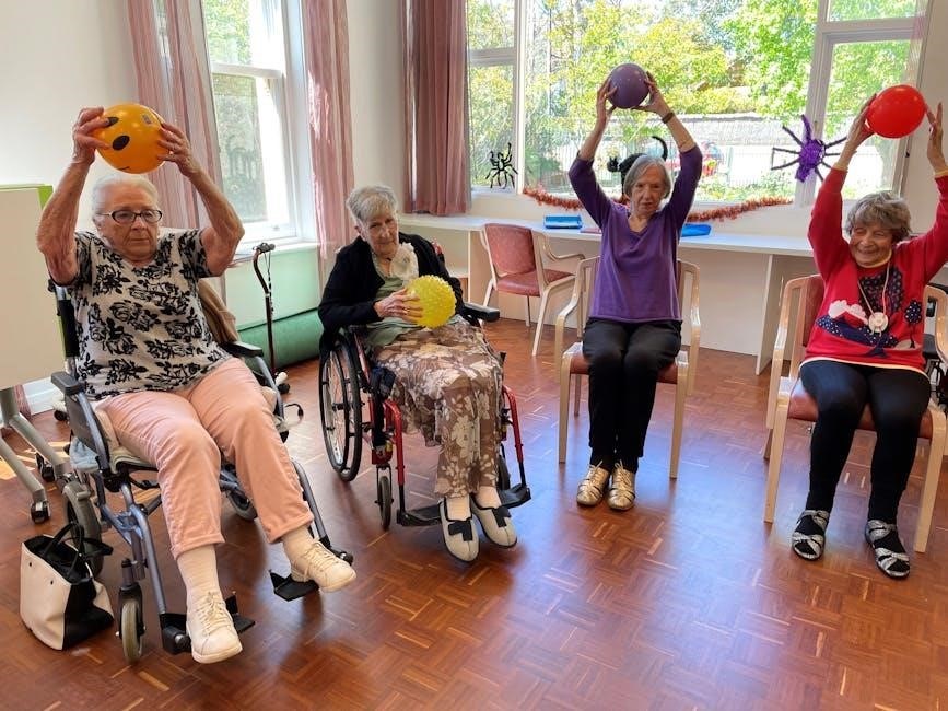
Proprioceptive Training
Restoring proprioception – the body’s awareness of its position in space – is crucial post-fracture. Begin with weight shifts in various directions, progressing to single-leg stance with minimal support. Utilize balance boards or foam pads to challenge stability. Incorporate exercises that mimic functional movements, like reaching and turning. Focus on controlled movements and maintaining balance. Proprioceptive deficits can contribute to re-injury, so consistent training is essential for regaining confidence and preventing falls during functional activities.
Advanced Rehabilitation Phase (6-12 Weeks)
This phase focuses on restoring full function and preparing for return to activity. Functional exercises, including gait training with varied terrain and stair negotiation, are prioritized. Advanced strengthening incorporates plyometrics – jump training – and progressive resistance training to build power and endurance. A multidisciplinary approach ensures proper form and progression. Return to activity considerations involve a gradual increase in intensity, guided by pain levels and functional assessments. The goal is to achieve independent mobility and participation in desired activities.

Functional Exercises – Gait Training, Stairs
Gait training progresses from simple ambulation to more complex patterns, addressing deviations and optimizing biomechanics. Initial focus is on proper weight-bearing and step length. Stair climbing is introduced gradually, starting with assisted ascents and descents, then progressing to independent performance. Emphasis is placed on quadriceps and gluteal activation for control. Proprioceptive exercises are integrated to enhance balance and coordination during functional movements. The aim is to restore a normal, pain-free gait pattern and independent stair negotiation.
Advanced Strengthening – Plyometrics, Resistance Training
This phase focuses on maximizing lower extremity strength and power. Resistance training utilizes progressive loading with exercises like squats, lunges, and hamstring curls. Plyometrics, including jump training and hopping, are introduced cautiously to improve explosive power and reactive muscle strength. Core stabilization exercises are crucial for maintaining proper form and preventing re-injury. The goal is to achieve strength symmetry between the injured and uninjured leg, preparing the patient for high-level activities and return to sport or demanding physical tasks.
Return to Activity Considerations
Returning to activity requires a gradual, individualized approach. Factors include fracture type, healing progress, and patient goals. A comprehensive assessment of strength, range of motion, and functional capacity is essential. Initial activities should be low-impact, progressively increasing in intensity and complexity. Monitoring for pain or swelling is crucial; any recurrence necessitates modifying the activity level. Consideration must be given to potential hardware limitations and long-term joint health. A successful return prioritizes safety and minimizes the risk of re-injury, ensuring sustained functional improvement.
Intramedullary Nailing Rehabilitation Specifics
Rehabilitation post-intramedullary nailing focuses on weight-bearing progression and addressing unique challenges. Protocols vary based on fracture location (proximal femur, shaft) and nail stability. Early mobilization is encouraged, but weight-bearing restrictions are initially enforced. Decision-making regarding weight-bearing is critical, guided by radiographic evidence of healing. Rehabilitation emphasizes regaining range of motion, strengthening surrounding musculature, and restoring proprioception. Potential complications, like delayed union, require modified protocols. A multidisciplinary approach optimizes outcomes following this common fixation method.
Weight-Bearing Protocols Post-Intramedullary Nailing

Weight-bearing progression after intramedullary nailing is carefully managed. Initial phases typically involve toe-touch weight-bearing, progressing to partial weight-bearing (PWB) with assistive devices. Full weight-bearing (FWB) is permitted based on radiographic evidence of callus formation and fracture stability. Protocols are individualized, considering fracture type, patient factors, and surgeon preference. Regular monitoring assesses pain levels and stability. Premature loading can compromise fixation, while delayed loading may hinder bone healing. Careful adherence to prescribed protocols is crucial for optimal recovery.
Rehabilitation Challenges with Intramedullary Nailing
Rehabilitation post-intramedullary nailing presents unique challenges. Early pain management and edema control are vital, alongside regaining range of motion. Muscle weakness, particularly in quadriceps and hamstrings, is common and requires targeted strengthening. Proprioceptive deficits necessitate specific training. Patients may experience discomfort around the nail insertion site. Addressing these issues requires a tailored exercise program, close monitoring, and a multidisciplinary approach involving surgeons and physiotherapists to optimize functional outcomes and minimize complications.
Complications and Considerations
Femur fracture rehabilitation can encounter complications. Non-union or delayed union requires prolonged immobilization and potential revision surgery. Hardware failure, though rare, necessitates surgical intervention. Managing pain and stiffness is crucial, often requiring multimodal approaches including medication and manual therapy. Vigilant monitoring for these issues is essential. Addressing these challenges promptly optimizes recovery. Careful consideration of individual patient factors, like pre-existing conditions, is vital for a successful rehabilitation process and preventing long-term disability.
Non-Union and Delayed Union
Non-union signifies a fracture’s failure to heal within expected timelines, while delayed union indicates slower-than-anticipated healing. These complications often necessitate prolonged immobilization, potentially with bone stimulation techniques. Physical therapy focuses on protecting the fracture site while maintaining surrounding muscle strength. Weight-bearing restrictions are strictly enforced. Surgical revision, including bone grafting, may become necessary. Addressing underlying factors like infection or inadequate blood supply is paramount. Careful monitoring of radiographic evidence guides treatment adjustments.
Hardware Failure
Hardware failure, encompassing plate breakage, screw loosening, or nail bending, demands immediate medical attention. Physical therapy’s role shifts to protecting the compromised fixation while minimizing further stress. Weight-bearing is typically restricted, and bracing may be implemented. Revision surgery is often required to restore stability. Post-operatively, rehabilitation progresses cautiously, prioritizing controlled loading and strengthening. Monitoring for signs of infection or implant migration is crucial. A multidisciplinary approach, involving orthopedic surgeons and therapists, ensures optimal outcomes.
Managing Pain and Stiffness
Persistent pain and stiffness post-femur fracture necessitate a multifaceted approach. Physical therapy utilizes modalities like heat, ice, and electrical stimulation alongside manual techniques to address soft tissue restrictions. Gentle range of motion exercises are crucial, progressing as tolerated. Pain management strategies include pharmacological interventions prescribed by the physician. Patient education on activity modification and self-management techniques is vital. Addressing psychological factors contributing to pain perception is also important for optimal functional recovery and improved quality of life.
Role of Multidisciplinary Team
Effective femur fracture rehabilitation demands a collaborative, multidisciplinary team. The orthopedic surgeon directs surgical intervention and monitors bone healing; Physiotherapists implement tailored exercise programs, focusing on range of motion, strengthening, and functional mobility. Rehabilitation specialists contribute expertise in maximizing functional independence and return to activity. Consistent communication between team members ensures coordinated care, addressing evolving patient needs. This integrated approach optimizes outcomes, minimizing complications and facilitating a successful recovery journey, enhancing the patient’s overall well-being.
Orthopedic Surgeon Collaboration
Close collaboration with the orthopedic surgeon is paramount throughout the rehabilitation process. The surgeon provides critical information regarding fracture stability, weight-bearing restrictions, and surgical findings. Regular communication ensures the physical therapy program aligns with the bone’s healing trajectory. Surgeons assess progress, modifying protocols as needed, and address potential complications like non-union or hardware issues. This partnership guarantees patient safety and optimizes functional outcomes, ensuring a coordinated and effective recovery following surgical fixation, particularly with intramedullary nailing.
Physiotherapist’s Role
The physiotherapist is central to restoring function after a femur fracture. They conduct comprehensive assessments, developing individualized exercise programs focusing on pain management, edema control, and range of motion. Early mobilization and proprioceptive training are key, alongside progressive strengthening of the quadriceps, hamstrings, and glutes. Physiotherapists educate patients on safe movement patterns, weight-bearing progression, and functional activities like gait training and stair negotiation. They monitor progress, adapting the program to maximize recovery and facilitate a return to pre-injury activity levels.
Rehabilitation Specialist Input
A rehabilitation specialist offers crucial expertise in complex cases. They provide advanced assessment of functional limitations and develop highly tailored rehabilitation plans, particularly post-intramedullary nailing. Their input is vital when addressing challenges like delayed union or non-union, optimizing weight-bearing protocols, and managing persistent pain or stiffness. They collaborate with the orthopedic surgeon and physiotherapist, ensuring a cohesive approach. Specialists can also address psychological aspects of recovery and facilitate a safe, effective return to activity, considering individual patient needs and goals.
Long-Term Follow-Up and Maintenance
Sustained commitment to strengthening and conditioning is paramount for long-term success. Continued exercise regimens should focus on maintaining quadriceps, hamstring, and gluteal strength, alongside proprioceptive training. Regular monitoring is essential to detect and address any late complications, such as hardware failure or developing stiffness. Proactive prevention of re-injury involves activity modification and adherence to a safe progression of functional tasks. Consistent follow-up appointments with the multidisciplinary team ensure optimal outcomes and sustained quality of life post-fracture.
Continued Strengthening and Conditioning
Maintaining gains requires a progressive, long-term exercise program. This includes consistent resistance training targeting the quadriceps, hamstrings, and gluteal muscles. Plyometric exercises, introduced cautiously, enhance power and functional capacity. Focus on core stability to improve overall biomechanics and reduce re-injury risk. Regular cardiovascular conditioning, like cycling or swimming, supports overall fitness. Adherence to a home exercise program, guided by the physiotherapist, is crucial for sustained improvement and independent function post-rehabilitation.
Preventing Re-Injury
Minimizing re-injury risk necessitates addressing biomechanical deficits and promoting safe movement patterns. Continued proprioceptive training enhances joint awareness and stability. Maintaining adequate muscle strength and flexibility is paramount. Gradual return to activity, avoiding sudden increases in load or intensity, is essential. Fall prevention strategies, particularly for older adults, should be implemented. Regular monitoring for pain or functional limitations allows for timely intervention. Adhering to a long-term conditioning program ensures sustained protection and optimal function.
Monitoring for Late Complications
Long-term follow-up is crucial for detecting potential late complications. Patients should be monitored for signs of non-union or delayed union, hardware failure, and persistent pain. Regular assessment of gait, range of motion, and functional abilities is essential. Vigilance for developing stiffness or limitations is key. Addressing any new symptoms promptly prevents progression. Periodic radiographic evaluation may be necessary to assess fracture healing and hardware integrity. Proactive management minimizes long-term disability and optimizes outcomes.