
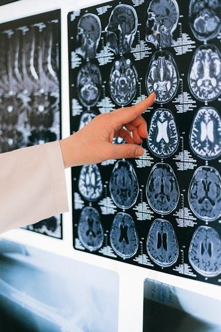
The human brain, a complex organ, orchestrates thought, emotion, and action. Understanding its structure—cerebrum, cerebellum, and brainstem—is key.
Detailed charts, often available as PDFs, visually map these parts and their functions, aiding comprehension of this vital organ’s intricacies.
Neuroanatomy reveals how each region contributes to cognition and behavior, while circulation diagrams highlight critical arteries like the MCA, ACA, PCA, and Basilar.
Overview of Brain Function
The brain’s primary function is to integrate sensory information and direct motor responses, serving as the central command center for the entire nervous system. This remarkable organ doesn’t just react; it learns, adapts, and shapes our individual identities. A comprehensive understanding begins with recognizing its three main divisions: the cerebrum, cerebellum, and brainstem, each uniquely contributing to overall functionality.
The cerebrum governs higher-level processes like thought and emotion, while the cerebellum focuses on coordination and balance. The brainstem, crucial for survival, manages basic life functions. Visualizing these components is greatly aided by brain parts charts, frequently available in PDF format for easy access and study.
These charts often detail key arteries—the Middle Cerebral Artery (MCA), Anterior Cerebral Artery (ACA), Posterior Cerebral Artery (PCA), and Basilar Artery— illustrating the brain’s circulatory network. Studying these resources provides a foundational understanding of how the brain regulates everything from breathing to complex cognitive tasks.
Importance of a Brain Parts Chart
A brain parts chart, particularly in PDF format, is an invaluable tool for anyone seeking to understand the complexities of the human brain. It provides a visual roadmap to navigate the intricate network of structures responsible for everything we think, feel, and do. These charts aren’t merely anatomical diagrams; they are essential for students, medical professionals, and anyone curious about neuroscience.
Detailed charts clearly delineate the cerebrum, cerebellum, and brainstem, alongside their respective lobes and key structures like the hippocampus and amygdala. They also illustrate critical circulatory components, such as the Middle, Anterior, Posterior Cerebral, and Basilar arteries, highlighting their role in brain health.
By visually connecting structure to function, these resources facilitate deeper learning and retention. Accessing these charts as PDFs allows for convenient study and reference, making the fascinating world of neuroanatomy more accessible to all.
The Three Main Parts of the Brain
The brain comprises three key sections: cerebrum, cerebellum, and brainstem. Charts visually demonstrate how each part contributes to vital functions, aiding comprehension.
PDF resources offer detailed views of these structures and their roles in controlling bodily functions and interpreting sensory input.
Cerebrum: The Largest Part
The cerebrum, the brain’s most substantial component, is responsible for higher-level functions like thought, language, and voluntary movement. Detailed brain charts, frequently available in PDF format, visually delineate the cerebrum’s complex structure and its crucial role in integrating sensory information.
These charts often showcase the four lobes – frontal, parietal, temporal, and occipital – each specializing in distinct cognitive processes. The frontal lobe governs executive functions, while the parietal lobe handles sensory processing. The temporal lobe is central to memory and audition, and the occipital lobe manages visual processing.
PDF resources provide a comprehensive understanding of the cerebrum’s intricate folds (gyri and sulci), increasing its surface area and, consequently, its processing capacity. Studying these charts aids in grasping how the cerebrum orchestrates complex behaviors and shapes individual identity. Understanding the cerebrum’s anatomy is fundamental to comprehending overall brain function.
Cerebellum: Coordination and Balance
The cerebellum, though smaller than the cerebrum, plays a vital role in coordinating movement, maintaining balance, and ensuring smooth, precise motor control. Brain parts charts, often accessible as downloadable PDFs, illustrate the cerebellum’s unique structure and its connections to other brain regions.
These charts highlight the cerebellum’s folded surface, maximizing its surface area within a limited space, and demonstrate its crucial role in refining motor commands initiated by the cerebrum. It doesn’t initiate movement, but perfects it.
PDF resources detailing neuroanatomy reveal how the cerebellum receives sensory input from the spinal cord and other brain areas, allowing it to adjust movements in real-time. Damage to the cerebellum results in impaired coordination and balance. Visual aids, like those found in PDF charts, are invaluable for understanding this complex interplay and appreciating the cerebellum’s contribution to fluid, graceful motion.
Brainstem: Basic Life Functions
The brainstem, a critical structure connecting the brain to the spinal cord, governs essential life-sustaining functions. Detailed brain parts charts, frequently available as PDF downloads, visually demonstrate the brainstem’s components – midbrain, pons, and medulla oblongata – and their interconnectedness.
These charts emphasize the brainstem’s role in regulating breathing, heart rate, blood pressure, and sleep-wake cycles. It acts as a relay station for sensory and motor information traveling between the brain and the body.
PDF resources focused on neuroanatomy illustrate how damage to the brainstem can have devastating consequences, impacting consciousness and even leading to death. Understanding its functions, aided by visual representations in PDF format, is crucial for appreciating its fundamental importance. Charts also show the location of key arteries supplying the brainstem, like the Basilar artery, vital for circulation.

Lobes of the Cerebrum

The cerebrum’s four lobes – frontal, parietal, temporal, and occipital – each specialize in distinct functions. PDF charts detail these areas and their roles in cognition.

Frontal Lobe: Executive Functions
The frontal lobe, the brain’s largest, governs higher-level cognitive processes collectively known as executive functions. These crucial abilities include planning, decision-making, working memory, and problem-solving – essentially, the skills that define our personality and allow for goal-directed behavior.
Detailed brain charts, frequently available in PDF format, visually delineate the frontal lobe’s subdivisions and their specific contributions. These charts often highlight areas responsible for motor control, speech production (Broca’s area), and complex thought.
Damage to the frontal lobe can result in significant behavioral and cognitive deficits, impacting impulse control, emotional regulation, and the ability to initiate tasks. Understanding the intricate functions of this lobe, aided by visual resources like anatomical PDFs, is paramount for neurological assessment and rehabilitation. The frontal lobe truly is the command center of the brain, orchestrating our conscious experience and guiding our interactions with the world.
Parietal Lobe: Sensory Processing
The parietal lobe plays a critical role in processing sensory information from across the body, integrating touch, temperature, pain, and spatial awareness. It’s responsible for our perception of location, navigation, and understanding our body’s position in space – essential for coordinated movement and interaction with the environment.
Comprehensive brain parts charts, often found as downloadable PDFs, illustrate the parietal lobe’s organization and its connections to other brain regions. These visual aids demonstrate how sensory input is mapped and interpreted, contributing to our overall perception of the world.
Damage to the parietal lobe can lead to difficulties with spatial orientation, recognizing objects, and even neglecting one side of the body. Studying detailed anatomical diagrams, particularly those in PDF format, is crucial for understanding the complexities of sensory processing and the impact of neurological injury on these vital functions.
Temporal Lobe: Memory and Audition
The temporal lobe is fundamentally involved in auditory processing, memory formation, and language comprehension. It receives and interprets sounds, enabling us to recognize voices and understand speech. Crucially, it houses the hippocampus, vital for forming long-term memories, and plays a role in emotional responses.
Detailed brain charts, frequently available as PDF resources, visually represent the temporal lobe’s intricate structures and their interconnectedness. These charts highlight the areas responsible for different types of memory – episodic, semantic, and procedural – and their relationship to auditory pathways.
Understanding the temporal lobe’s functions is essential for comprehending conditions like amnesia or auditory processing disorders. Accessible PDF diagrams provide a valuable tool for students and professionals alike, offering a clear depiction of this complex brain region and its critical role in cognition and behavior.
Occipital Lobe: Visual Processing
The occipital lobe is the visual processing center of the brain, responsible for interpreting information from the eyes. It receives signals related to shape, color, and motion, allowing us to perceive the world around us. Damage to this lobe can result in various visual impairments, ranging from mild distortions to complete blindness.
Comprehensive brain parts charts, often distributed as PDF documents, provide a detailed anatomical view of the occipital lobe and its subdivisions. These charts illustrate the pathways visual information travels through, from the retina to the visual cortex, and highlight areas specialized for different aspects of vision.
Utilizing these visual aids enhances understanding of how the brain constructs our visual reality. They are invaluable resources for students, medical professionals, and anyone seeking to learn more about the complexities of visual perception and the brain’s remarkable capabilities.

Detailed Brain Structures & Their Functions
Exploring structures like the hippocampus and amygdala reveals crucial roles in memory and emotion. PDF charts visually detail these areas, enhancing comprehension.
Hippocampus: Memory Formation
The hippocampus, a critical structure within the brain, plays a pivotal role in the formation of new memories. Often depicted in detailed brain charts – readily available as PDFs – it’s essential for converting short-term memories into long-term storage.
These charts visually demonstrate the hippocampus’s location within the temporal lobe, highlighting its connections to other brain regions involved in memory processing. Damage to this area can result in significant memory impairments, particularly the inability to form new declarative memories – facts and events.
Understanding the hippocampus’s function is crucial for comprehending cognitive processes. PDF resources often illustrate how the hippocampus works in conjunction with the amygdala (emotional processing) and other cortical areas to consolidate and retrieve memories. Studying these charts provides a valuable insight into the complexities of human memory and the brain’s remarkable capacity for learning and recall.
Amygdala: Emotional Processing
The amygdala, a key limbic system component, is fundamentally involved in processing emotions, particularly fear and aggression. Detailed brain parts charts, frequently found as downloadable PDFs, clearly illustrate its location deep within the temporal lobes, showcasing its connections to other crucial brain areas.
These visual aids demonstrate how the amygdala rapidly assesses threats, triggering physiological responses like increased heart rate and heightened alertness. It’s not solely about negative emotions; the amygdala also plays a role in experiencing positive emotions and attaching emotional significance to memories.
PDF resources often depict the amygdala’s interplay with the hippocampus, highlighting how emotional experiences enhance memory consolidation. Understanding the amygdala’s function is vital for comprehending emotional regulation, anxiety disorders, and the brain’s overall response to environmental stimuli. Charts provide a valuable visual guide to this complex emotional center.
Thalamus: Sensory Relay Center
The thalamus functions as the brain’s central relay station for sensory information. Comprehensive brain parts charts, often available in PDF format, visually demonstrate its strategic location, positioned deep within the brain to receive input from various sensory systems – excluding smell – before forwarding it to the cerebral cortex for further processing.
These charts illustrate how nearly all incoming sensory signals (vision, hearing, touch, taste) pass through the thalamus, acting as a crucial filter and organizer. It doesn’t just passively relay; the thalamus also modulates sensory signals, influencing attention and consciousness.
PDF resources often highlight the thalamus’s connections to both the cerebral cortex and the brainstem, showcasing its role in regulating sleep, alertness, and motor control. Understanding the thalamus is essential for grasping how the brain integrates and interprets the world around us, and charts provide a clear visual representation of its complex connectivity.

Brain Circulation and Key Arteries
Brain charts, often in PDF form, detail vital arteries like the MCA, ACA, PCA, and Basilar. These diagrams illustrate blood flow, crucial for oxygenating brain tissue.
Understanding arterial pathways is key to comprehending potential impacts of circulatory disruptions on brain function.
Middle Cerebral Artery (MCA)
The Middle Cerebral Artery (MCA) is a major player in cerebral circulation, prominently featured in brain anatomy charts and PDF resources. It supplies a significant portion of the lateral cerebral hemispheres, impacting extensive areas responsible for crucial functions.
Specifically, the MCA irrigates regions governing motor and sensory functions for the face, arm, and hand, as well as speech and language comprehension – often depicted clearly in detailed neurovascular maps. Occlusion of the MCA, frequently illustrated in medical diagrams, can lead to contralateral hemiparesis (weakness on one side of the body), sensory loss, and aphasia (language difficulties).
PDF charts often highlight the MCA’s branching pattern, demonstrating its supply to the frontal and parietal lobes. Understanding its territory is vital for interpreting neurological deficits and planning treatment strategies. Visual aids, like those found in Hamilton Health Sciences resources, are invaluable for grasping the MCA’s anatomical significance and clinical implications.
Anterior Cerebral Artery (ACA)
The Anterior Cerebral Artery (ACA) is a critical component of the brain’s circulatory system, frequently detailed in anatomical charts and comprehensive PDF guides. It provides blood supply to the medial aspects of the frontal and parietal lobes, areas crucial for higher-level cognitive functions and motor control.
Specifically, the ACA nourishes regions responsible for leg and foot motor function, as well as areas involved in personality, executive functions, and behavioral control – often visually represented in neurovascular diagrams. Blockage of the ACA, commonly illustrated in medical resources, can result in contralateral leg weakness, behavioral changes, and difficulties with executive decision-making.
PDF charts often showcase the ACA’s characteristic branching pattern, highlighting its supply to the medial frontal lobe. Understanding its territory is essential for interpreting neurological symptoms and guiding clinical interventions. Resources like those from Hamilton Health Sciences provide valuable visual aids for grasping the ACA’s anatomical importance and potential clinical consequences.
Posterior Cerebral Artery (PCA)
The Posterior Cerebral Artery (PCA) is a major artery supplying blood to the occipital lobe, a region vital for visual processing, and portions of the temporal lobe. Detailed anatomical charts, frequently available as downloadable PDFs, clearly illustrate the PCA’s course and branching pattern.
These resources demonstrate the PCA’s role in nourishing areas responsible for visual cortex function, including processing color, motion, and form. Occlusion of the PCA, often depicted in medical illustrations, can lead to visual field deficits – specifically, homonymous hemianopia – impacting one side of the visual field in both eyes.
PDF guides also highlight the PCA’s supply to parts of the thalamus and midbrain, influencing sensory relay and motor control. Understanding the PCA’s territory is crucial for diagnosing neurological conditions and interpreting neuroimaging findings. Charts from sources like Hamilton Health Sciences provide a clear visual representation of its anatomical significance and clinical implications.
Basilar Artery
The Basilar Artery, formed by the vertebral arteries, is a critical vessel supplying the brainstem and cerebellum with oxygenated blood. Comprehensive brain circulation charts, often found as downloadable PDF resources, meticulously detail its path and branching structure.
These visual guides illustrate how the Basilar Artery feeds crucial areas governing essential life functions, including breathing, heart rate, and consciousness. Blockage of the Basilar Artery, frequently depicted in medical diagrams, can result in severe neurological deficits, including locked-in syndrome.
PDF anatomical references also showcase the Basilar Artery’s contribution to the posterior cerebral circulation, ultimately supplying the occipital lobes. Charts from institutions like Hamilton Health Sciences emphasize its clinical relevance in stroke diagnosis and treatment; Understanding the Basilar Artery’s anatomy is paramount for neurovascular assessment and intervention.

Resources for Further Learning (PDF Charts)
Numerous online resources offer detailed PDF charts illustrating the intricate anatomy of the human brain. Hamilton Health Sciences provides valuable diagrams focusing on brain function and circulation, specifically detailing arteries like the Middle, Anterior, Posterior Cerebral, and Basilar arteries.
These downloadable charts are invaluable for students, medical professionals, and anyone seeking a deeper understanding of neuroanatomy. They visually represent the cerebrum, cerebellum, and brainstem, alongside the four lobes – frontal, temporal, parietal, and occipital.
Searching for “brain parts and functions chart PDF” yields a wealth of options, including resources outlining the hippocampus, amygdala, and thalamus. These visual aids enhance comprehension of complex neurological concepts, offering a clear pathway to learning about the brain’s remarkable structure and function.